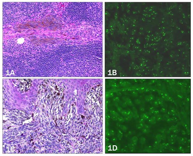
Figure 1. Case 1, 64 year-old white male with lymph node involvement (a,b) originating from a primary melanoma, 1.0 mm in thickness on the right upper arm (c,d).
a) Pigmented, spindled melanocytes within trabecula of the sentinel lymph node (10× objective). b) Increased copy number of 11q13 (CCND1) by FISH as evidence by more than two green signals per nucleus (40x objective). c) Primary melanoma (20× objective). d) Primary melanoma with similarly increased copy number of 11q13 (40× objective).

