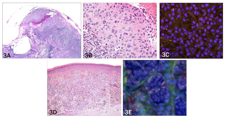
Figure 3. Case 6 need to put in description as in 1 and 4.
a) Collections of epithelioid melanocytes in the lymph node capsule, extensively involving the lymph node circumferentially (2× objective). b) Melanocyte nuclei show only mild variation in shape and are approximately three times the size of adjacent lymphocytes (40 × objective). c) Primary tumor with sheets of monomophic melanocytes with moderate cytologic atypia (10× objective), and d) scattered deep mitoses (20× objective).

