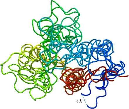
Figure 4.

Snapshots from the denatured ensemble depicting motions of the protein backbone. The structures are colored from red at the N terminus (looking down the helix axis) to blue at the C terminus. The 2-, 2.5-, 3-, 3.5-, and 4-ns structures are displayed. The 6-Å separation labeled in the figure is shown merely to provide perspective.
