Figure 2.
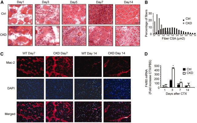
CKD suppresses muscle regeneration in vivo induced by injuring muscle to activate satellite cells. (A) Representative hematoxylin- and eosin-stained cross-cryosections of injured TA muscles were obtained from control and CKD mice. Regenerated myofibers were identified by their central nuclei. Injured muscles from CKD mice had increased infiltration of cells and slowed regeneration. Bar = 20 μm. (B) At 1 month after injury, fibers were immunostained for laminin, and the sizes of myofibers with central nuclei were measured. The histogram of the sizes of the myofibers with central nuclei indicates that CKD impaired regeneration. (C) At days 7 and 14 after injury, muscles were immunostained for macrophages (Mac-2 antibody; red) and DAPI for nuclei (blue). At both times, macrophages were increased in muscles of CKD mice. Bar = 50 μm. (D) Sequential changes in F4/80 mRNAs after CTX injury were detected by RT-PCR and normalized for 18S mRNA. The fold increase over PBS treatment confirmed that macrophages were more plentiful in muscles of CKD mice. (*P < 0.05 versus results in injured muscles of control mice; n = 5 mice in each group).

