
Figure 3.
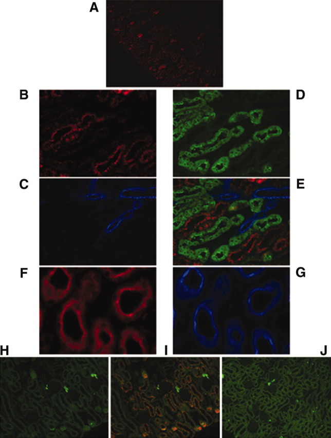
Atrap protein in the mouse kidney localizes predominantly to the proximal tubule. (A) Atrap immunostaining in the renal cortex of a wild-type mouse with marked staining of the proximal tubule. (B through D) Higher magnification pictures of co-staining with anti-Atrap (red; B) anti-SM22 (blue, as a marker for the renal vasculature; C), and anti-calbindin (green, as a marker for the distal convolute tubule; D). (E) An overlay. (F and G) Staining of cross-sections of the proximal tubule with anti Atrap (red; F) and anti megalin (blue; G). (H through J) Immunostaining for renin (green; H) and co-staining with anti-Atrap (red) in the renal cortex of an Atrap+/+ (I) and Atrap−/− (J) mouse.