
Figure 4.
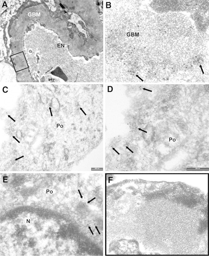
AR is present in GBM and in subepithelial immune deposits. (A and B) AR is present in GBM as shown by several scattered gold particles in the context of electron-dense subepithelial immune deposits (arrows). (C through E) AR was also observed in the cytoplasm and the plasma membrane of podocyte bodies (Po). (E) AR is not expressed at the nuclear (N) level; instead, AR is present in the cytoplasm as indicated by arrows. (F) Blank sample lacking primary antibody was totally negative (see also Supplemental Figure 5, A, C, and E). EN, endothelium. Magnifications: ×8900 in A; ×56,000 in B, D, and E; ×36,000 in C; ×44,000 in F.