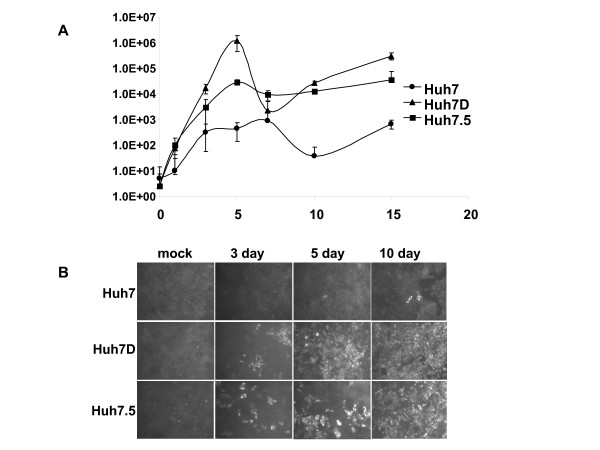
Figure 3.
A. Growth of HCV2a J6/JFH1 in Huh7, Huh7D, and Huh7.5 cells. The indicated cells were mock infected or infected the J6/JFH1 strain of HCV at an m.o.i. of 0.01. After 6 hours, cells were washed with growth medium three times and passed to 12 well plates. Cells were collected at the indicated time points and frozen at -70°C. Virus was tittered as described in the text. Ffu, focus forming units. Error bars represent the standard error. B. Growth of HCV2a J6/JFH1 in Huh7, Huh7D, and Huh7.5 cells assessed by IF. The indicated cells were mock infected or infected the J6/JFH1 strain of HCV at an m.o.i. of 0.01. After 6 hours, cells were washed with growth medium for three times and passed to 96 well plates. HCV antigen was detected at the indicated time points by immunfluorscence.

