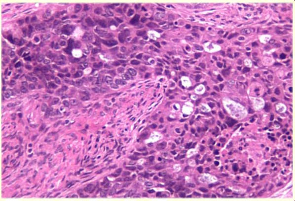
Figure 3.

Representative section of ovary stained with hematoxylin and eosin (100×) demonstrating irregular nests of large anaplastic cells with an ill-defined cribriform architecture infiltrating the ovary. No residual normal ovarian tissue is shown.
