Figure 1.
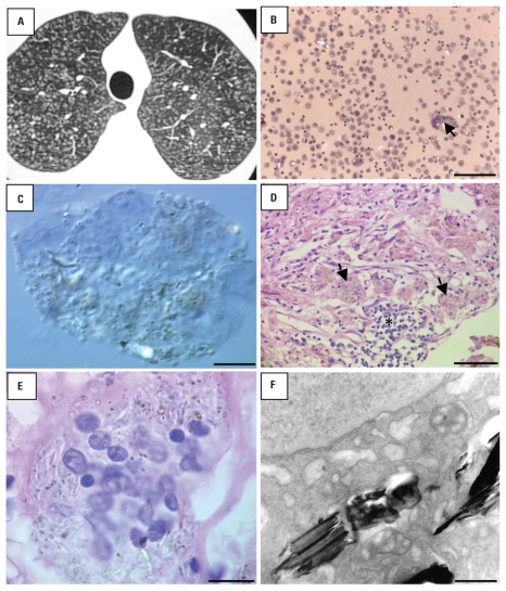
Images resulting from various tests performed on the patient. (A) HRCT image showing small nodular opacities throughout lung fields. (B) Bright-field microscopy image of BALF showing numerous macrophages and a multinucleated giant cell (arrow) containing bright refractile particles; bar = 100 μm. (C) Differential interferential contrast microscopy of a multinucleated giant cell in BALF with refractile particles within the cytoplasm; bar = 5 μm. (D) Light microscopy of the lung biopsy specimen (hematoxylin and eosin stain) showing many foreign-body–type granulomas (asterisk) and giant cells (black arrows); bar = 100 μm. (E) Detail of a multinucleated giant cell with numerous round and elongated particles within the cytoplasm (hematoxylin and eosin stain); bar = 7.5 μm. (F) TEM image of a macrophage showing platelike materials within the cytoplasm; bar = 0.25 μm.

