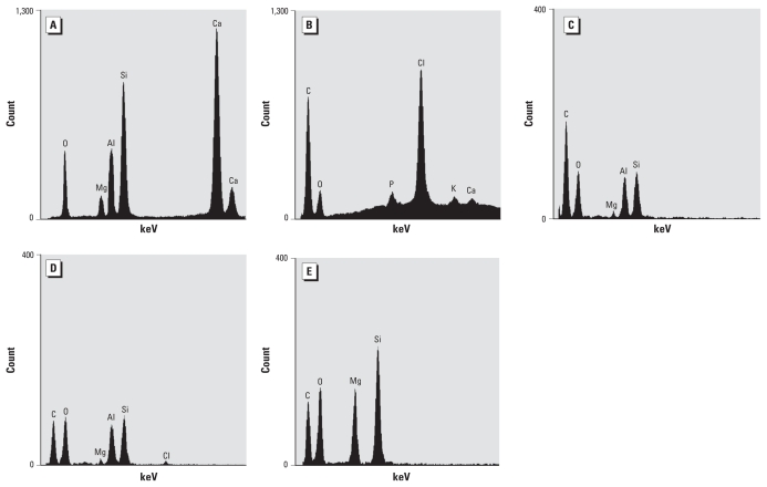
Figure 2.
EDXA spectra obtained by TEM. (A) EDXA spectrum obtained from the fibers brought in by the patient. (B) Spectrum from the resin also used in the lamination process. (C) Spectrum from a platelike material in the patient’s alveolar macrophages derived from the lung biopsy; this spectrum is compatible with kaolinite. (D) Spectrum of a different platelike material in the lung alveolar macrophage, suggesting the presence of resin because of Cl. (E) Spectrum from an amorphous material in the lung alveolar macrophages indicating a talc-like material.

