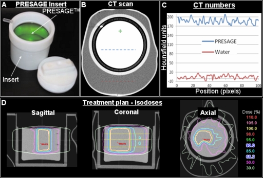
Figure 2.
Treatment planning for the RPC phantom containing the customized Presage 3D dosimetry insert. (a) A photograph of the customized insert. (b) Central slice of the x-ray CT scan of the combined phantom. (c) A plot of CT numbers along dotted lines in B. (d) Isodose distribution in sagittal, coronal, and axial planes. The same treatment plan used to irradiate the RPC phantom with the standard insert was also used to irradiate the phantom with Presage insert with the exception that the prescription dose was reduced from 6.6 to 4 Gy to avoid over exposing the dosimeter.

