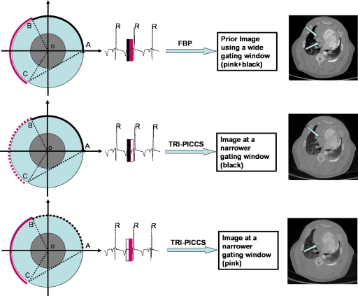
Figure 2.
Illustration of the proposed temporal resolution improvement using PICCS (TRI-PICCS) method. A standard FBP image reconstruction method is used to reconstruct a prior image from a short-scan data set acquired within a wide cardiac window (labeled by two narrow solid bars in the top panel of the figure). In the second step, only half of the short-scan data is used to reconstruct two separate cardiac images using the new TRI-PICCS algorithm. In each case, the short-scan reconstruction was used as the prior image. The resulting images (middle right and lower right) were reconstructed using cardiac windows of one-half the width. The reconstructed images clearly demonstrate how the motion artifacts in the prior image (arrows in the upper right image) are eliminated when the cardiac window is reduced by a factor of 2. This effectively improves the temporal resolution by a factor of approximately 2.

