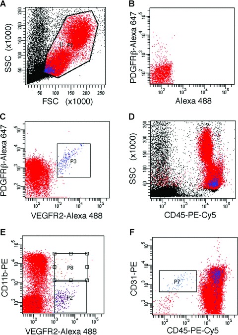
Figure 3.

Phenotypic characterization of CPCs. LSR-II flow cytometric analyses of (gated, a) mononuclear cells in peripheral blood, identified a distinct population of CPCs that co-express VEGFR2 and PDGFRβ (c); single-colour controls were used for compensation (e.g. see staining for PDGFRβ-Alexa Fluor 647 in b). These CPCs (in blue) are positive for CD45 (d) and show scattering properties typical of small cells with a high nucleus to cytoplasm ratio (a, d). A subset of CPCs is positive for CD11b (e), whereas all VEGFR2+PDGFRβ+ CPCs are positive for CD31 (f); CPCs are distinct from cells with a phenotype consistent with ‘circulating ECs’, i.e. CD31+CD45− (see gated population in f).
