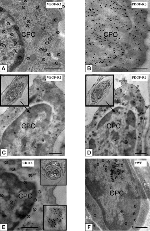
Figure 4.

Phenotypic characterization of CPCs in lungs post-HALI. Antigenic sites, visualized with 10-nm protein A-gold by high-resolution microscopy, demonstrated that CPCs were VEGFR2+ (a) and PDGFRβ+ (b) post-HALI (weeks 6 and 7). Representative images of the same CPC profile in adjacent sections of lung tissue (c and d, and insets) demonstrated co-expression of VEGFR2 and PDGFRβ antigenic sites post-HALI (week 7). Other representative images of additional antigenic sites expressed by CPCs post-HALI (week 7): CD11b (e and inset) and for vWF (f). Typically, the sites for CD11b were uniformly distributed over CPCs but also appeared as clusters (e, see bottom inset, illustrating an example of a cluster of ∼40 CD11b+ labelled sites). Ten-nm gold-labelled antigenic sites (a, c–f) are circled for clarity to distinguish these from 7 to 8 nm ribosomes on cell profiles. Ninety-nm-thick Unicryl resin sections stained with uranyl acetate and lead citrate. Bars = 0.5 μm (a–f).
