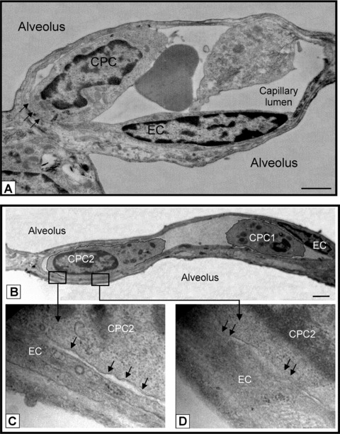
Figure 5.

CPCs contact and adhere to endothelium post HALI. (a) Representative image of CPC aligning and adhering to a capillary EC (see arrows) post-HALI (week 7). At this time, approximately 35% of CPCs form contacts with lung capillary ECs. (b) Representative image of two CPCs in a lung capillary post-HALI (week 6): CPC1 appears free in the lumen and CPC2 is in the process of adhering to the adjacent EC (see boxed areas): higher magnification shows regional separation of two distinct cell membranes (lower left image, arrows), and regional loss of membrane delineation between the two cells (lower right image, arrows). Eighty-nm-thick epon resin sections stained with uranyl acetate and lead citrate. Bars = 1 μm (a) and 2 μm (b).
