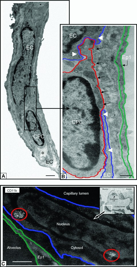
Figure 6.

CD11b+ CPCs and cells integrate into capillary surfaces post HALI. Illustrations of CD11b+ CPC (a) and capillary EC in a residual capillary structure post-HALI (week 6). An asterisk marks the restricted capillary lumen at this time. A region of the CPC cytosol is inserted between the processes of two capillary ECs (b, black arrows). Note the location of the CPC within the capillary lumen in relation to the processes of adjacent ECs (white arrowheads), to (an un-delineated) perivascular cell process, and to the underlying epithelial (Ep1) cell process. The blue line delineates the plasmalemmal membrane of the capillary EC, the red line outlines the CPC, and the green line demarcates the adjacent epithelial type 1 (Ep1). (c) Representative image of lung capillary (see boxed area of inset) and higher magnification of the luminal surface illustrating cell clusters of antigenic sites for CD11b (circled in red) typical of CPCs expressing this protein post-HALI (week 7). Image is inverted to highlight the location of this cell with antigenic sites. The other EC indicated in the same capillary (see inset, small arrow) does not express CD11b. Eighty-nm-thick epon resin section (a, b), and 90-nm-thick Unicryl resin section (c), stained with uranyl acetate and lead citrate. Bars = 1 μm (a), 0.5 μm (b, c).
