
Figure 1.
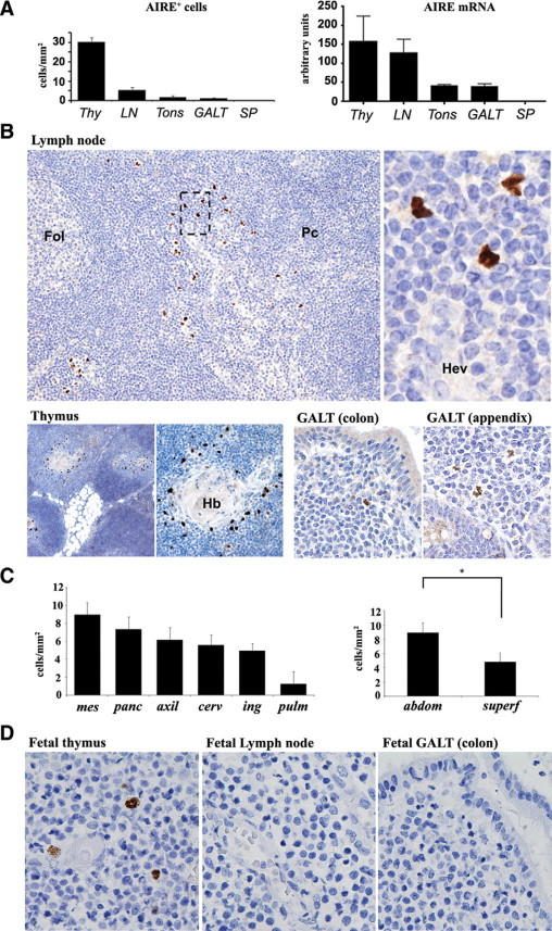
Expression of AIRE in human thymus and peripheral lymphoid tissues. A: The number of AIRE+ cells/mm2 evaluated by immunohistochemistry (left plot) and AIRE mRNA levels analyzed by quantitative RT-PCR (right plot) show that extrathymic AIRE expression occurs particularly in LNs, albeit at lower level than in the thymus (Thy), tonsils (Tons), and GALT but not in the spleen (SP). B: In LNs (top panel) AIRE+ cells are distributed as scattered cells in the interfollicular area/outer paracortex (Fol, follicle; Pc, paracortex), in the vicinity of high endothelial venules (Hev), as indicated in the higher magnification from the dotted box area (original magnification: ×10 and ×80). In the thymus (bottom panel, left), AIRE+ cells occur in the medulla and correspond to medullary epithelial cells around Hassall's bodies (Hb) (original magnification: ×4 and ×10). Rare cells are present in the colon, appendix, and GALT (bottom panel, right) (original magnification: ×20). C: The number of nodal AIRE+ cells differs in anatomical regions (left plot). The difference between AIRE+ cells in abdominal (mesenteric plus peripancreatic) compared with superficial (axillary, inguinal, and cervical) LNs is statistically significant (right plot; *P < 0.001). mes, mesenteric; panc, peripancreatic; axil, axillary; cerv, cervical; ing, inguinal; pulm, pulmonary; abdom, abdominal; superf, superficial. D: In the fetus, only the thymus contains AIRE+ cells in the medulla, whereas no positive cells occur in LNs and GALT (all panels, original magnification: ×40). All immunostains for AIRE were performed using diaminobenzidine/hydrogen peroxide as the chromogen and hematoxylin as the nuclear counterstain. Bar graphs show averages and SEs of at least three independent measurements.