
Figure 2.
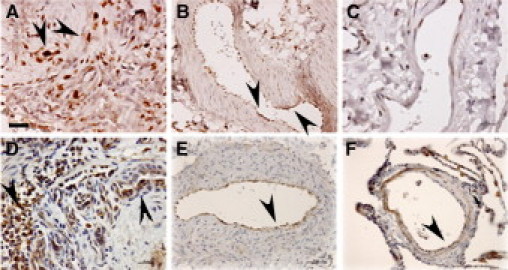
Expression of HIF-1α and its transcriptional target CAIX in IPAH plexiform lesions and remodeled pulmonary arteries. Note increased expression of HIF-1α in cells forming the vascular slits in IPAH plexiform lesions (A, arrowheads) and intimal cells lining the remodeled pulmonary arteries (B, arrowheads), when compared with intima of a normal pulmonary artery (C). This pattern of HIF-1α expression correlated with expression of its transcriptional target CAIX in an IPAH plexiform lesion (D, arrowheads) and pulmonary artery intima (E, arrowhead) when compared with a normal pulmonary artery (F, arrowhead). Scale bars: 25 μm (A, D, E); 50 μm (B, C, F). Images representative of overall five IPAH and five normal lungs tested.