
Figure 2.
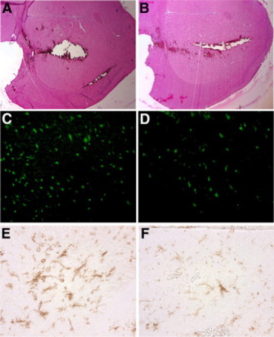
Histological consequences of ICH and the effect of minocycline applied in the blood that was used to create injury. Photomicrographs A to F show the histopathology of brain injury 24 hours after ICH. The area of brain damage (A and B; H&E stain), FJ-B positive dying neurons (C and D), and activity of microglia/macrophages (E and F) are reduced by 10 μg/ml of minocycline (B, D, and F) mixed within the blood that was used to create ICH compared with PBS vehicle (A, C, and E). Original magnification: ×400 (C–F); ×25 (A and B).