
Figure 3.
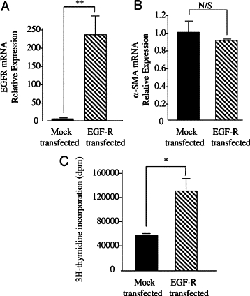
Overexpression of EGF-R in aged dermal fibroblasts. Aged dermal fibroblasts were transfected with either EGF-R-pCR3.1 (EGF-R transfected) or GFP (mock transfected). Transfection efficiency of EGF-R-pCR3.1 was 41.5%. Total mRNA was extracted and EGF-R (A) and α-SMA (B) mRNA expression was assessed by RT-Q-PCR. Ribosomal RNA expression was used as an endogenous control, and gene expression was assessed relative to mock transfected. The comparative CT method was used for relative quantification of gene expression, and the results represent the mean ± SE of nine individual experiments using cells isolated from three different donors. Statistical analysis was performed by Student's t-test: **P < 0.01. N/S, not significant. C: EGF-induced proliferation was determined by incorporation of [3H]thymidine. Expression plasmids for EGF-R or GFP (mock transfected) were introduced into aged fibroblasts using Nucleofector technology before metabolic labeling, performed by incubation with 1 μCi/ml d-[3H]thymidine after addition of either serum-free medium alone (black bars) or serum-free medium containing 10 ng/ml EGF (gray bars) for 24 hours. Data are the mean ± SE from six individual experiments, using cells isolated from two different donors. Statistical analysis was performed by Student's t-test: *P < 0.05.