
Figure 1.
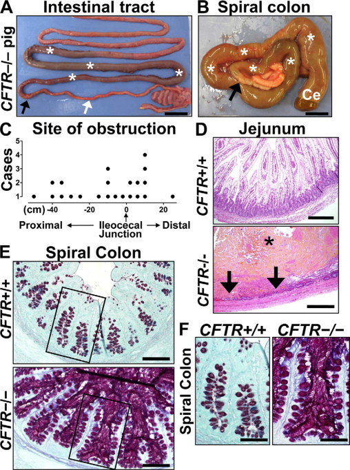
Meconium ileus in CFTR−/− intestine. A: Meconium-filled bowel (asterisks) was proximal to the obstruction interface (black arrow); distal to the interface, the bowel was hypoplastic (white arrow). Scale bar = 2.45 cm. B: Meconium (white asterisks) dilated the spiral colon (ventral view) and cecum (Ce) proximal to the obstruction interface (black arrow). Microcolon appeared distally. Scale bar = 1.34 cm. C: Sites of obstruction in CFTR−/− pigs were distributed on either side of the ileocecal junction extending from the small intestine (proximal) into the spiral colon (distal). D:CFTR−/− jejunum showing luminal meconium (asterisk) causing severe distension and mucosal thinning with focal hemorrhage, necrosis (arrows), and neutrophilic inflammation, HE stain. Scale bars = 0.29 mm. E:CFTR−/− spiral colon had luminal mucus accumulation with hyperplastic mucus-producing cells especially noticeable compared with CFTR+/+ along the surface epithelium (inset boxes, see F). ABPY stain. Scale bars = 117 μm. F:CFTR−/− spiral colon glands were distended by stringy mucus that extends from stout mucus-producing cells in the epithelium, ABPY stain. Scale bars = 69 μm.