
Figure 3.
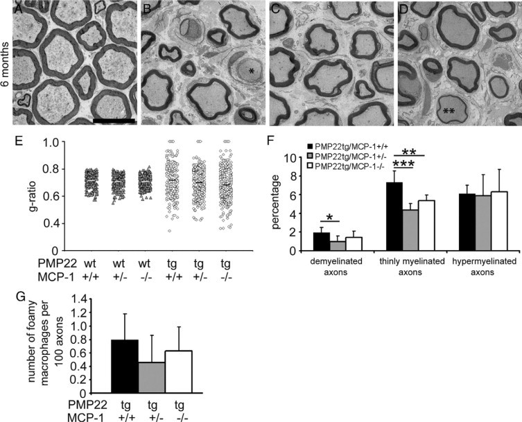
Demyelination is ameliorated in peripheral nerves of PMP22tg/MCP-1+/− and PMP22tg/MCP-1−/− mice. Morphometric analysis of femoral nerves from wild-type and PMP22 mutant mice (n = 3 to 4). Electron microscopy of femoral quadriceps nerves of six-month-old PMP22wt/MCP-1+/+ (A), PMP22tg/MCP-1+/+ (B), PMP22tg/MCP-1+/− (C), and PMP22tg/MCP-1−/− mutants (D). Analysis of femoral quadriceps nerves revealed fewer features indicative of demyelination in PMP22tg/MCP-1+/− and PMP22tg/MCP-1−/− mice, such as demyelinated axons (asterisk) and thinly myelinated axons (double asterisk). Scale bar = 10 μm. E: G-ratios reflect the milder demyelinating neuropathy in nerves of PMP22tg/MCP-1 double mutant mice. F: Quantification of pathological alterations confirms the ameliorated demyelination in femoral quadriceps nerves of PMP22tg/MCP-1+/− and PMP22tg/MCP-1−/− mice, whereas hypermyelination is independent from MCP-1/CCL2-expression. G: Foamy macrophages containing myelin debris showed a tendency toward lower numbers in femoral quadriceps nerves of PMP22tg/MCP-1+/− mice compared with nerves of PMP22tg/MCP-1+/+ and PMP22tg/MCP-1−/− mice. *P < 0.05, **P < 0.01, ***P < 0.001, Mann–Whitney U test.