
Figure 3.
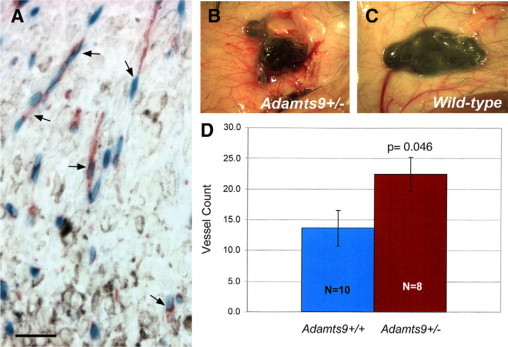
ADAMTS9 is expressed in tumor capillaries and influences the induction of tumoral vessels. A: Section through the periphery of a B.16-F10 melanoma implanted in ADAMTS9+/− mice shows that ADAMTS9 (β-gal staining, blue) is expressed by capillary endothelial cells (arrows) (red, endomucin) invading the melanoma (pigmented cells). Scale bar = 50 μm. B, C: Representative images of a B.16-F10 melanoma in ADAMTS9+/− mice (B) and wild-type (C) mice showing the enhanced vasculature around the tumor in ADAMTS9+/− mice. D: Quantitative analysis of in vivo angiogenic activity induced by B.16-F10 melanomas in ADAMTS9+/− and wild-type mice. Mice were sacrificed 9 days after intradermal injection of tumor cells. Radially oriented blood vessels entering the periphery of the tumors were counted by an observed blinded to the genotype of the mice. The mean vessel count ± the SEM is shown. The P value is shown.