
Figure 6.
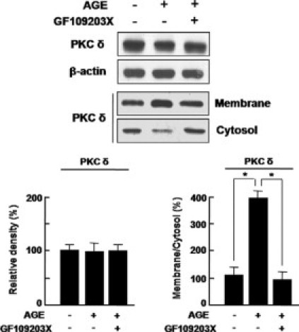
Translocation of PKC δ from cytosol to membrane in HRMECs was significantly increased under diabetic condition, but not PKC δ expression. HRMECs were incubated for 12 hours with or without a pan-PKC inhibitor, GF109203X (5 μmol/L) in AGE treatment. Cell extracts were fractionated as a membrane and a cytosol fraction. In each fraction, PKC δ expression was assessed by Western blotting. β-Actin served as the loading control. Figures were selected as representative data from three independent experiments. Quantitative analysis was performed by measuring protein expression relative to the control. Each point represents the mean (±SD) of three independent experiments, each performed in triplicate. *P < 0.05.