Figure 4.
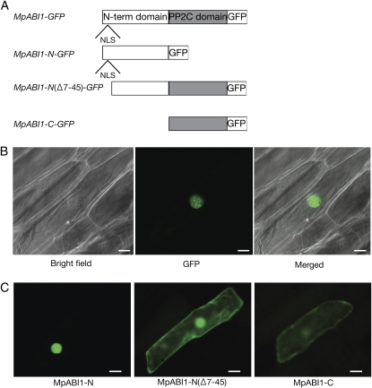
Localization study using GFP fusion proteins. A, Schematic representation of GFP fusion constructs of MpABI1 and its mutant constructs used for analysis. B, Cellular localization of MpABI1-GFP. The DNA construct for the MpABI1-GFP fusion protein was bombarded into epidermal cells of onion, and the cells were observed after 1 d of incubation at 25°C in the dark. Bright-field, GFP fluorescence, and merged images are shown. Bars = 30 μ m. C, N-terminal domain-directed nuclear localization of MpABI1. Fluorescent images show onion epidermal cells bombarded with DNA constructs for the MpABI1 N-terminal domain (MpABI1-N; amino acids 1–225), MpABI1-N (Δ 7–45) lacking NLS, and the C-terminal PP2C domain (MpABI1-C; amino acids 213–568) fused to the upstream region of the GFP coding sequence. Bars = 50 μ m.

