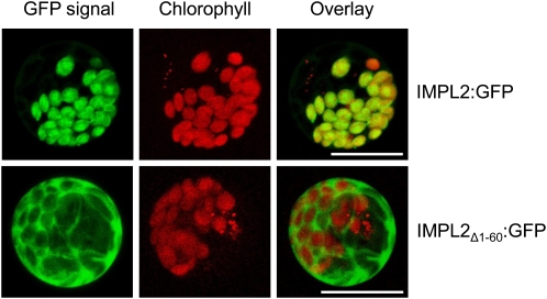
Figure 7.
Subcellular localization of IMPL2. Confocal images of maize protoplasts transiently expressing either full-length IMPL2 protein fused to GFP (IMPL2:GFP) or a truncated IMPL2 protein lacking the first 60 amino acids predicted to act as the plastid transit peptide fused to GFP (IMPL2Δ 1–60:GFP). GFP emission at 500 to 520 nm (GFP signal), chlorophyll autofluoresence at 575 to 650 nm (chlorophyll), and a merged image (overlay) are shown. Scale bars indicate a distance of 20 μ m.

