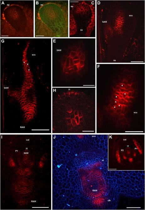
Figure 4.
ZmPIN1 protein localization during maize embryogenesis. All images portray maize B73 embryo sections labeled with the anti-AtPIN1 antibody plus a secondary antibody carrying the Alexa568 fluorochrome. G to I and K present laser confocal images, while A to F and J present epifluorescence images acquired with a Leica DC300F camera. Green and blue color in B and J represents tissue autofluorescence collected with GFP and DAPI filters, respectively. E and F represent bigger magnification of D, while H reports a detail of K. During the proembryo stage, ZmPIN1 proteins mark a group of cells placed in the middle of the proembryo, without any evident polarity, while no signal is detectable in the suspensor (A and B). In the early transition phase, ZmPIN1 proteins are expressed in the upper cells of the proembryo and detectable and polarized only in the anticlinal membranes of differentiating embryo protoderm, indicating an auxin flux directed upward and converging toward the embryo tip (C). Later on, at the transition stage, ZmPIN1 proteins mark the initials of the SAM and inner tissues of the developing scutellum (D). The SAM initials are characterized by a central group of cells presenting apolar ZmPIN1 protein localization (E), while along the scutellum, the proteins mark the apical cell membranes, indicating the presence of an acropetal auxin flux directed toward the tip of the scutellum (F). Arrowheads in F indicate the polarization of ZmPIN1 proteins in apical cell membranes. At the late coleoptilar stage, an inversion of ZmPIN1 polarization is evident in the scutellum: after scutellum vasculature differentiation, the ZmPIN1 proteins are localized in the basal cell membranes, suggesting an auxin flux directed downward (G). Arrowheads in G indicate the polarization of ZmPIN1 proteins in basal cell plasma membranes. ZmPIN1 protein relocation at the level of the scutellum corresponds to the establishment of a continuous basally polarized localization of the ZmPIN1 proteins at the level of embryo axis (G). Therefore, two basipetal auxin fluxes can be detected at this stage: one from the scutellum tip to the SAM and RAM, and another from the SAM toward the root pole (G and I). Maize SAM initiates up to six true leaf primordia prior to seed dormancy, and during these processes, ZmPIN1s localize in all the cell membranes of the central group of cells in the SAM, while in the L1 outer layer ZmPIN1s localize only at the level of the incipient primordia (H, I, and K). ZmPIN1 proteins are basally localized in primordia provasculature, suggesting auxin fluxes directed from the primordia to the inner tissues of the SAM and then to the root pole (I and K). ZmPIN1 protein polarization also suggests basipetal auxin transport from the SAM to the RAM (I and J), and the proteins are also detectable in the coleorhiza (I and J) and at the sites of seminal root initiation (J). Moreover, when coleoptile is fully developed, a longitudinal section shows that ZmPIN1 proteins mark the apical cell membranes, suggesting auxin fluxes directed toward the coleoptile tip (K). clh, Coleorhiza; col, coleoptile; ep, embryo proper; I1, incipient primordium; P1 and P2, leaf primordia; scu, scutellum; sr, seminal root; su, suspensor. Bars = 100 μ m in D, G, I, and J; 75 μ m in K; 50 μ m in A to C, E, and F; and 20 μ m in H.

