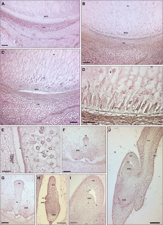
Figure 5.
IAA localization during maize kernel development. An anti-IAA monoclonal antibody was employed to determine the auxin distribution and accumulation in developing maize endosperm and embryos. IAA accumulation is detectable in the BETL domain at 8 DAP (B) and 12 DAP (C and D), in conductive cells at 12 DAP (C), and in the chalazal maternal tissues from 6 to 16 DAP (A–C), while only a weak signal is visible in the starchy endosperm (B, C, and E). Free IAA accumulation is detectable in ESR cells (F and G) and in the aleurone layer (E). Embryos at the globular stage are characterized by high auxin accumulation at the top of the embryo proper, while a very low signal is detectable in the suspensor (F). At the early and late transition stages, the auxin accumulation is evident in the protodermal layer and inner cells of the embryo proper, whereas suspensor shows the IAA minimum (G). The coleoptilar stage embryos are characterized by gradients of IAA distribution: the signal is high in the tip of the scutellum and in all the protoderm cells at the adaxial side, while it decreases toward the suspensor (H). Auxin maxima are detectable at the tip of the developing coleoptile, in the SAM, and in embryo root pole (H). During the SAM morphogenetic phase, auxin accumulation is evident in the tip of leaf primordia (I and J), in the corpus of the SAM (I and J), and in the RAM (J). al, Aleurone; CC, conductive cells; cha, chalaza; col, coleoptile; ep, embryo proper; I1, incipient primordium; P1, leaf primordium; pro, protoderm; scu, scutellum; se, starchy endosperm; su, suspensor. Bars = 100 μ m in A to C, F to H, and J; and 50 μ m in D, E, and I.

