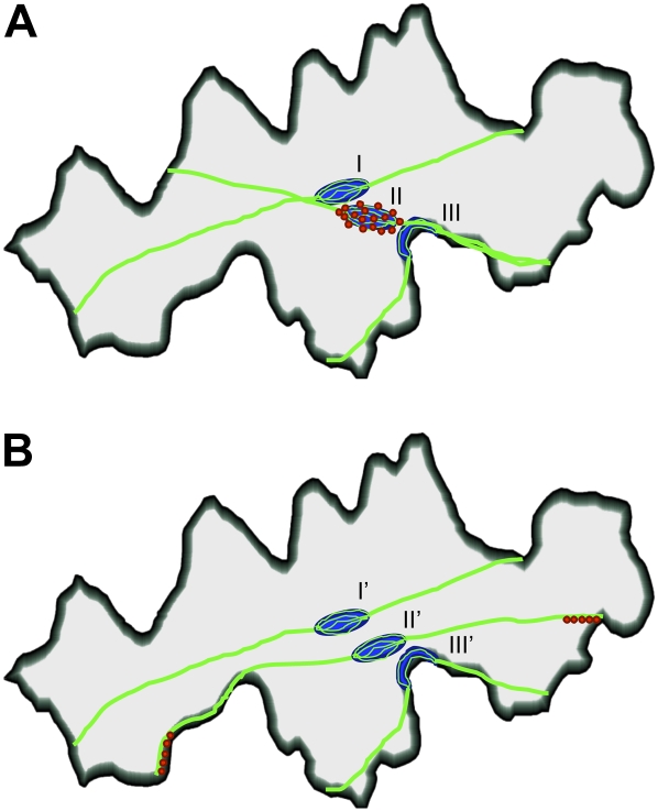
Figure 8.
Models for light-dependent nuclear motility in epidermal cells. A, In darkness, the nucleus (blue) is located at the cell center (along the inner cell wall) in association with thick, longitudinally arranged actin bundles (green; I). Under blue light, the bundles associated with the nucleus become connected to the anticlinal wall nearest the nucleus (II). The nucleus moves along the actin bundles and becomes anchored at the anticlinal wall (III). B, In darkness, the nucleus is located as in A (I'). Under blue light, actin bundles associated with the nucleus laterally move toward the anticlinal wall (II'), thereby drawing nucleus with them, where it eventually becomes anchored at the anticlinal wall nearest the center of cell (III'). Note that myosin (red) is specifically localized on the nuclear periphery and around the attachment site of the actin bundle to the anticlinal wall in models A and B, respectively.

