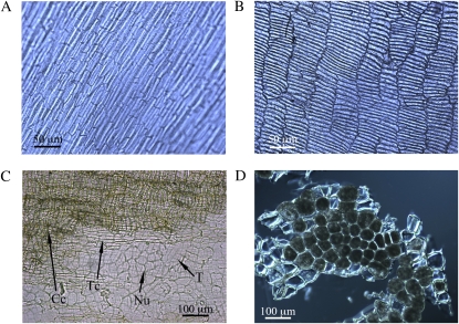
Figure 1.
Micrographs of the isolated bran fractions. A, Outer bran fraction (epidermis and hypodermis). B, Intermediate bran fraction (cross cells, tube cells, testa, and nucellar tissue). C, Detailed view of the individual layers in the intermediate fraction (Cc, cross cells; Nu, nucellar tissue; T, testa; Tc, tube cells). D, Aleurone cells. [See online article for color version of this figure.]

