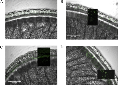
Figure 4.
Fluorescence immunolocalization of defense proteins in bran cross-sections overlaid on differential interference contrast images of cross-sections (labeled). Dark inset overlays in images show fluorescence labeling without the differential interference contrast overlay. A, Control treated only with secondary antibody. B, OXO antibody. C, XIP-1 antibody. D, PR-4 antibody.

