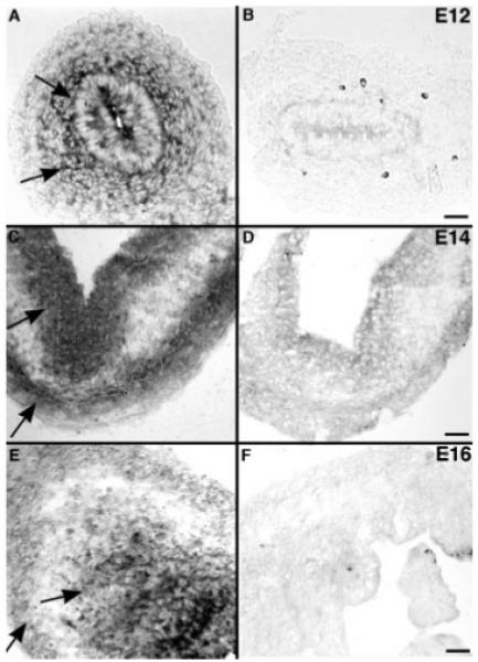
Figure 2.

Transcripts encoding laminin-111 chains in the fetal gut are located by in situ hybridization. (A) Gut (E12). Transcripts encoding laminin β1 are detected in the endoderm and surrounding mesenchyme (arrows). The expression of laminin β1 is most intense closest to the epithelium and decreases in a centrifugal concentration gradient; few cells in the outer gut mesenchyme express laminin β1. (B) Gut (E12). No labeling is detected with the sense probe. (C) Gut (E14). Transcripts encoding laminin β1 are found in two distinct zones, the endoderm and the outer gut mesenchyme (arrows), without an intervening concentration gradient. (D) Gut (E14). No labeling is detected with the sense probe. (E). Gut (E16). Transcripts encoding laminin α1 are found in distribution similar to that of transcripts endoding β1 at E14; cells expressing mRNA encoding laminin α1 (arrows) are found in both the endoderm and the outer gut mesenchyme. (F) Gut (E16). No labeling is detected with the sense probe. Scale bars = 50 μm in A, B; 25 μm in C–F.
