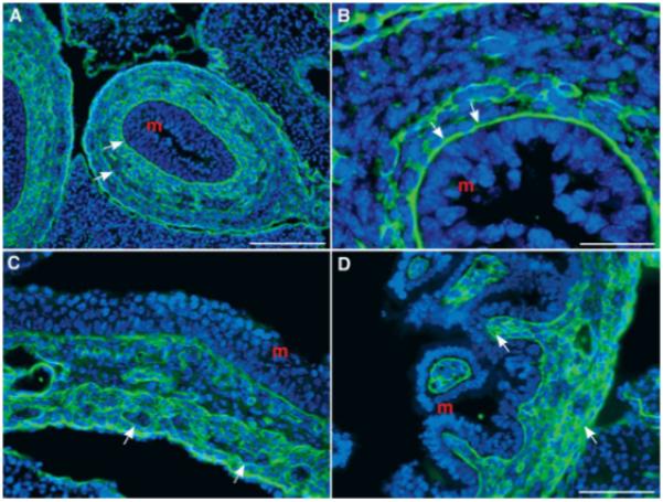
Figure 3.

Laminin immunoreactivity is found in the fetal mouse gut. (A) Colon (E13). Laminin immunoreactivity is concentrated in the basal laminae and is also found outlining cells of the mesenchyme (arrows; m = mucosa). The pattern of immunoreactivity reveals a concentration gradient that is greatest near the mucosa and is similar to that seen with in situ hybridization [see Fig. 2(A)]. (B) Colon (E13). The same section as A at higher magnification. The concentration of laminin immunoreactivity is clearly seen in the basal laminae (arrows; m = mucosa). (C) Stomach (E15). Laminin is still concentrated near the mucosa, but is now also detected in the outer gut mesenchyme. The ganglia of the myenteric plexus are outlined by laminin and can be detected by their negative image because of the absence of intraganglionic laminin immunoreactivity (arrows; m = mucosa). (D) Small bowel (E15). Laminin immunoreactivity is abundant in the gut wall and, as in C, is concentrated near the mucosa and in the outer gut mesenchyme (arrows; m = mucosa). Scale bars = 100 μm in A; 25 μm in B; 50 μm in C (applied to D).
