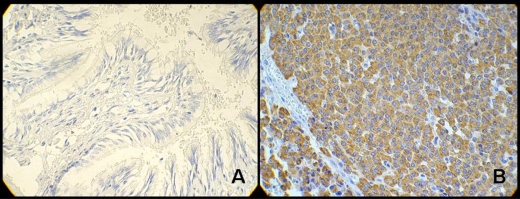
Figure 3.
Heparanase expression in the diagnosis of broncopulmonar carcinoid tumors. Optical microscopy at X400 power: A) negative expression of heparanase (absence of staining—peroxidase—in cell’s cytoplasm) in bronchial mucosa not compromised by neoplasm; B) positive expression of heparanase (presence of cytoplasm full of peroxidase—brownish areas) in broncopulmonar carcinoid tumor. (Adapted from: de Matos et al87).

