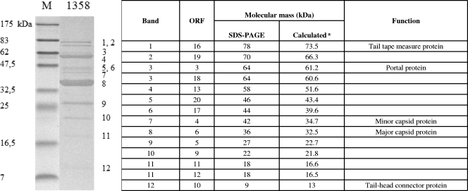
FIG. 4.
LC-MS/MS analysis of phage 1358 structural proteins. (Left) Coomassie blue staining of a 12% SDS-polyacrylamide gel showing phage 1358 structural proteins. Letters on the right indicate bands cut out of the gel and identified by LC-MS/MS. The sizes (in kDa) of the proteins in the broad-range molecular mass standard (M) are indicated on the left. (Right) Identification of phage 1358 proteins from corresponding bands shown in the left panel. Numbers at the right correspond to the numbers indicated in the left panel. aCalculated from the gene sequence.

