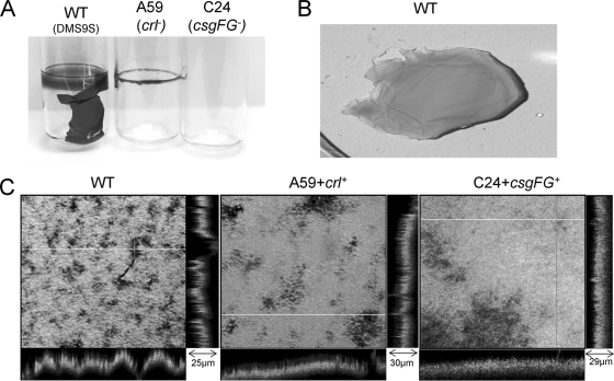
FIG. 8.
Pellicle formation at the air-liquid interface. Bacteria were incubated in LBNS for 3 days at 26°C in glass tubes, and their pellicles were visualized by staining with CV (A). The wt pellicle was removed from the tube and stained with AO (B). AO-stained pellicles of the wt and complemented mutants were examined by confocal microscopy (C). Stacks were collected and processed using FluoView 500 software. The z-section values of representative areas are denoted. Pictures were processed using Microsoft Office Picture Manager software to enhance their clarity.

