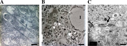
FIG. 4.
Ultrastructural evidence of hfq mutant invasion deficiency and the lack of differentiation observed using electron microscopy. (A) WT cells differentiated into bacteroids (scale bar, 1 μm). (B) hfq mutant (Smblφ500)-inoculated plants exhibit nodules with cells invaded (I) or not invaded (NI) by bacteria (scale bar, 10 μm). Noninvaded cells contain large granules of starch (S). Panel C shows a dividing cell failing to differentiate into a bacteroid (designated by an arrow) (scale bar, 1.5 μm).

