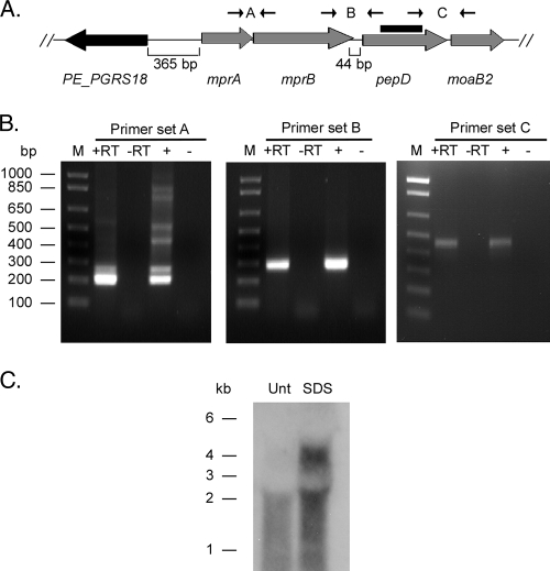
FIG. 2.
Transcriptional organization of the pepD locus in M. tuberculosis. (A) Chromosomal organization of pepD and surrounding genes in M. tuberculosis H37Rv. The primers used to determine the operon structure and the probe generated for Northern blot hybridization are shown. (B) Agarose gels depicting RT-PCRs with primer sets spanning intergenic regions. Lanes: M, marker lane; +RT, RNA treated with reverse transcriptase; −RT, RNA treated without reverse transcriptase; +, genomic DNA; −, no template. (C) Northern blot of RNA samples obtained from either untreated (Unt) or SDS-treated M. tuberculosis H37Rv cultures.

