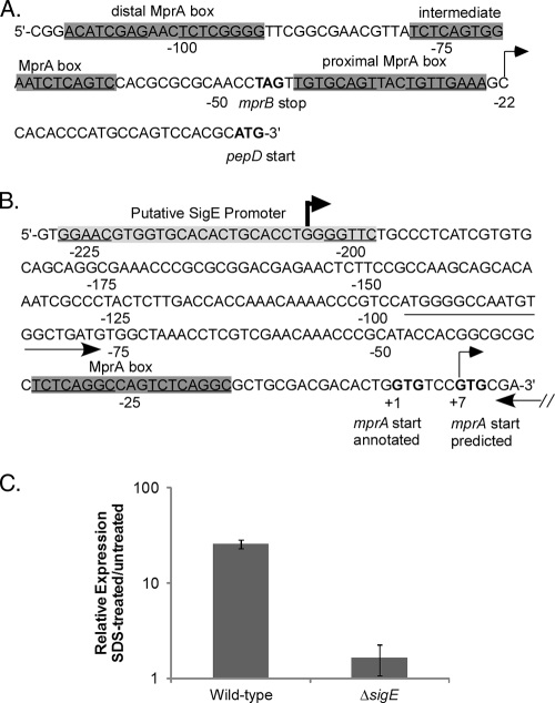
FIG. 3.
Characterization of the pepD promoter region. (A and B) Genomic depiction of pepD (A) and mprA (B) upstream regions, including MprA binding sites (gray boxes), determined transcriptional start sites (arrows), and annotated or predicted translational start and stop sites (boldface letters). Note that one of the transcriptional start sites for mprA is downstream of the translational start site previously annotated. The location of the primer set used to measure expression from the distal mprA promoter is also shown by arrows. (C) The wild type and the ΔsigE M. tuberculosis mutant were grown in the absence of SDS or were exposed to 0.05% SDS for 90 min, and the relative amounts of expression from the distal mprA promoter were determined. Expression levels were normalized to those of the 16S ribosomal gene, rrs, and are expressed as relative expression from cultures exposed to SDS versus untreated cultures. The results represent the means ± standard errors of the mean (SEM) from three independent experiments performed in triplicate.

