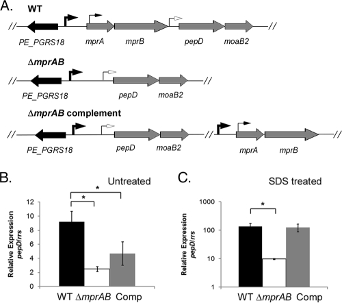
FIG. 5.
Polarity resulting from the ΔmprAB mutation. (A) Genomic organization of the mprA-mprB-pepD-moaB2 locus in M. tuberculosis H37Rv (wild type [WT], the ΔmprAB mutant, and complemented strains. The arrows depict the identified transcriptional start sites in the region. (B and C) Wild-type M. tuberculosis, the ΔmprAB mutant, and the ΔmprAB complemented strain were grown without SDS (B) or were exposed to 0.05% SDS for 90 min (C). The relative amounts of pepD in the strains were compared. The expression levels were normalized to that of the 16S ribosomal gene, rrs. The results represent the means ± SEM from three independent experiments performed in triplicate. *, P < 0.05.

