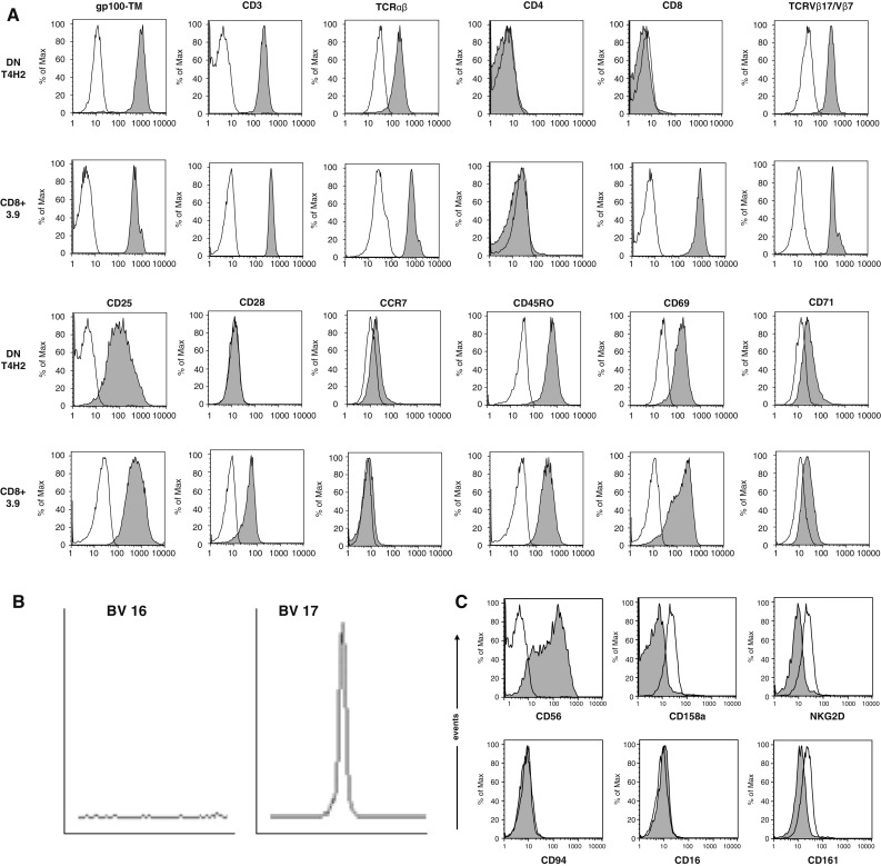
Fig. 1.

Phenotype of the gp100-reactive DN T cell clone T4H2. T4H2 cells were isolated from a melanoma patient after gp100-peptide vaccination and expanded over several months by repetitive stimulation with allogeneic feeder cells. A classical gp100-reactive CD8+ CTL clone (3.9), isolated from an HLA-A2+ healthy donor, served as a control. a T4H2 and 3.9 cells were harvested and stained with a panel of mAbs recognizing CD3, CD4, CD8, TCRαβ, TCRVβ17 (for T4H2) or TCRVβ7 (for 3.9), CD28, CD45RO, CCR7, CD69, CD25, CD71 and the HLA-A2/gp100-tetramer and analyzed by flow cytometry. Histograms are shown on gated lymphocytes (by forward and side scatter) to exclude residual feeder cells and fluorescence intensity is presented by the gray line. Open histograms show the respective isotype controls. b Analysis of the TCRVβ family distribution in T4H2 DN T cells via the TCR CDR3 spectratyping method. A clonal T cell population is represented by a distinct peak in the appropriate TCRVβ17 receptor family. One negative (TCRVβ16) and the TCRVβ17 positive run off results are shown. c Analysis of NK cell marker expression on T4H2 cells. T4H2 cells were stained with mAbs against NK cell markers CD56, CD158a, NKG2D, CD94, CD16, CD161, and analyzed by flow cytometry
