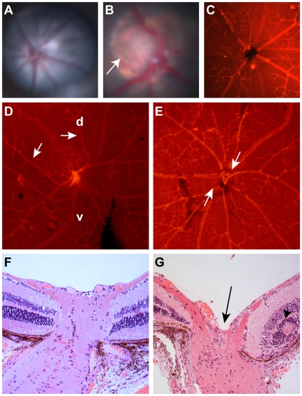
Figure 1. Clinical ocular phenotype in C57BL/6-Pax2+/A220G mice compared to wild-type, C57BL/6 mice.
(A) Fundus photograph of C57BL/6 mouse showing normal optic nerve and radial pattern of retinal blood vessels. (B) Fundus photograph of C57BL/6-Pax2+/A220G mouse showing congenital excavation of the optic nerve head with peripapillary pigment changes (arrow). (C) Lectin immunofluorescence of wild-type C57BL/6 mouse showing normal, radial vessel patterning. (D,E) Lectin immunofluorescence of C57BL/6-Pax2+/A220G mice showing abnormal vascular patterning, including curving of vessels towards the dorsal retina (D, arrows, d = dorsal, v = ventral) and separation of the central retinal vascular trunks (E, arrows). Histologic section of a Pax2+/+ (F) and a Pax2+/A220G (G) mouse eye through the optic nerve and peripapillary retina showing abnormal excavation of the optic nerve (G, arrow) and retinal rosette formation (G, arrowhead). Remnants of the tunica vasculosis lentis and mild extension of the retinal pigment epithelium were variably noted in histopathology from other Pax2A220G/+ eyes (data not shown).

