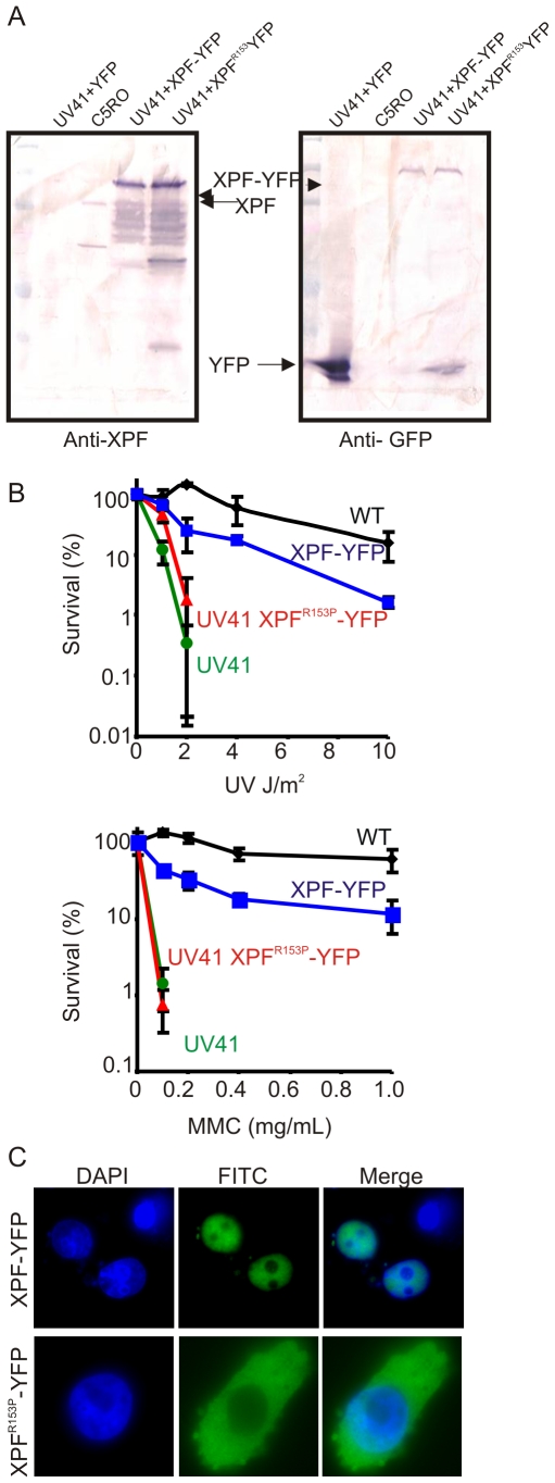
Figure 3. Characterization of XPF-YFP and XPF153-YFP in CHO cells.
(A) Western blot analysis of XPF-YFP expressed in Xpf mutant cells. XPF-deficient hamster cell line, UV41, was transiently transfected with wild type XPF-YFP or XPF153-YFP and the fusion proteins were detected using an antibody against XPF or GFP. C5RO was used as positive control for the XPF blot and as a negative control for the GFP blot. UV41 cells transfected with YFP alone was used as a negative control for XPF blot and as a positive control for GFP blot. (B) Clonogenic survival of wild-type (wt), XPF-deficient CHO cell line UV41, and UV41 transfected with wild type XPF-YFP and XPF153-YFP after UV and MMC treatment. Colonies were counted 7–10 days after treatment and results are plotted as mean 3 independent experiments. (C) Subcellular localization of wild type XPF-YFP and XPF153-YFP after transient transfection in XPF-deficient the CHO cell line UV41 detected by fluorescence microscopy.

