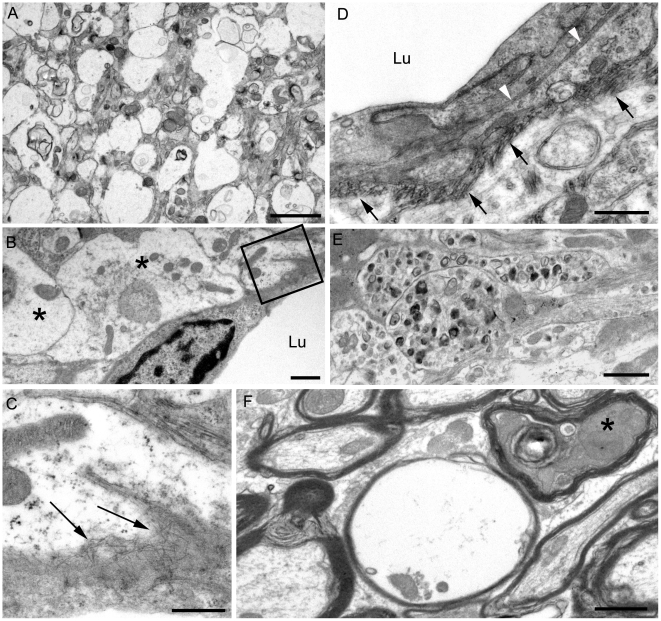
Figure 6. Ultrastructure of cerebral cortex and cerebellum of a 22L scrapie-infected tg44+/+ mouse.
Sections were from same mouse as shown in Figure 5, and were stained with uranyl acetate/lead citrate. (A) Area of neuropil with severe vacuolation where most vacuoles originate within processes and are separated from each other by intact membranes. (B) Several distended astrocytic processes (asterisks) of the perivascular glial limitans are present around a blood vessel Lu: lumen. (C) On higher magnification of boxed area from (B), the endothelial basement is shown to be filled with irregularly orientated amyloid fibrils (arrows). (D) The earliest stage of vascular amyloid is shown. Here the endothelial basement membrane (black arrowheads) is intact, but the pericyte basement membrane (black arrows) is thickened and heavily infiltrated with short amyloid fibrils (white arrowheads). (E) Severe neuritic dystrophy in which several processes show an excessive accumulation of organelles and abnormal electron dense bodies. (F) White matter of the cerebellum showing an empty myelin sheath (seen as vacuoles by light microscopy) and also a dark degenerate axon (asterisk) within an intact myelin sheath. Similar findings were observed in RML scrapie-infected tg44+/+ mice. Scale bars: A, 2 µm; B, E, F, 1 µm; C, 500 nm; D, 500 nm.

