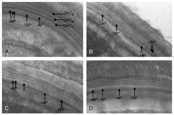
FIG. 2.
Light micrographs showing the orientation of hair cells in the cochlear anomaly in guinea pigs. First-row OHCs (OHC1), second-row OHCs (OHC2), and third-row OHCs (OHC3). Surface view of organ of Corti of a control guinea pig showing abnormal OHCs (arrow-heads), particularly in the OHC1. The rotation of OHC bundles can be in the 180-, +90-, −90-, +45-, and −45-degree direction.

