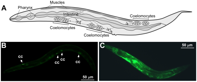
Figure 5. Visualizing the coelomocyte deficiency.
A schematic drawing of a worm shows that fluids secreted into the pseudocoelom from surrounding tissues accumulate in coelomocytes (A, modified from Fares and Greenwald, 2001 [18]). Epifluorescence micrographs of GS1912 (B) and NP717 (C) worms. In GS1912, ssGFP is expressed in body wall muscles from myo-3 promoter, secreted into the pseudocoelom and accumulated in coelomocytes [18]. White arrows indicate accumulation of GFP in coelomocytes (cc, B). As a result of coelomocytes ablation in NP717, GFP accumulates in the pseudocoelom [18] (C).

