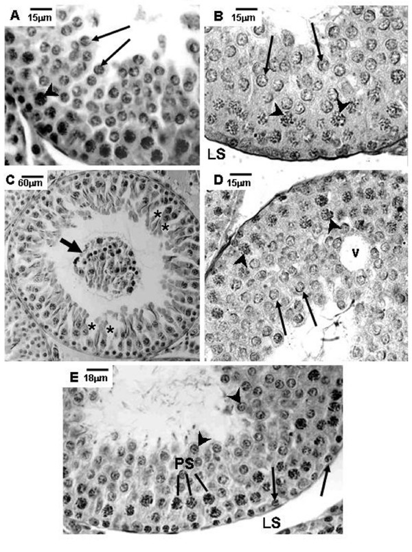
Figure 1.
Photomicrographs of testicular sections of Sham Control, Amifostine, Doxorubicin and Amifostine/Doxorubicin treated rats at different ages. PAS+H method. Portions of tubular sections of 45-day-old rats. A and B: Sham Control (A) and Amifostine-treated (B) groups showing organized seminiferous epithelium containing various cell types until round spermatids. Lymphatic space (LS); primary spermatocytes (arrowheads); round spermatids (long arrows). C and D: Doxorubicin-treated group. Note in C the cellular debris and the germinal lineage cells detached from the epithelium into the tubular lumen (short arrow); see also the discontinuous seminiferous epithelium (asterisks). In D, observe the vacuole formation (v), the primary spermatocytes (arrowheads) and the round spermatids (long arrows). E: Amifostine/Doxorubicin-treated group showing the organized seminiferous epithelium with normal morphology. Spermatogonia (arrows); primary spermatocytes (PS); round spermatids (arrowheads).

