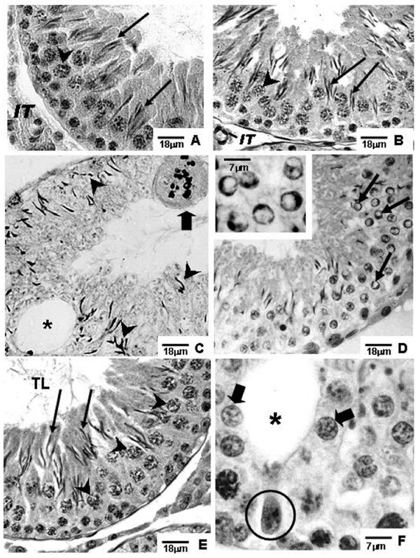
Figure 2.
Photomicrographs of testicular sections of Sham Control, Amifostine, Doxorubicin and Amifostine/Doxorubicin treated rats at different ages. PAS+H method. Portions of sections of seminiferous tubules of 60-day-old rats. A and B: Sham Control (A) and Amifostine (B) groups. Elongated spermatids (arrows); interstitial tissue (IT); primary spermatocytes (arrowheads). C and D: Doxorubicin-treated group displaying damaged seminiferous epithelium. In C, an accentuated epithelial depletion and various elongated spermatids, sometimes abnormally located (arrowheads) can be observed; vacuole (asterisk); multinucleated formation in degeneration (thick arrow). In D, some spermatids showing outlined condensed and ring-shaped marginal chromatin are observed; this event suggests apoptosis occurrence (thin arrows). Inset: detail of round spermatids with ring-shaped marginal chromatin. E and F: Amifostine/Doxorubicin-treated group. Note the presence of morphologically normal round (thick arrows) and elongated (long arrows) spermatids, vacuole (asterisk) and Sertoli cell nucleus dislocated from the tubular periphery and with abnormal chromatin condensation (circle); primary spermatocytes (arrowheads); tubular lumen (TL).

