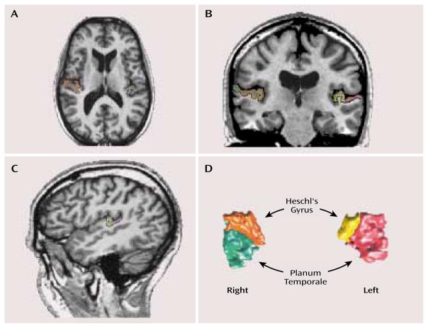
FIGURE 1.
Delineation of Heschl’s Gyrus and the Planum Temporale in the Brain of a Subject With Schizotypal Personality Disordera
a The axial image (part A) illustrates the most inferior slice boundary for Heschl’s gyrus, and the coronal image (part B) shows the separation of Heschl’s gyrus and the planum temporale. The sagittal view (part C) was used to confirm the delineation of the two regions of interest, which are shown in a three-dimensional rendering in part D. Following radiologic convention, the left side of the brain is on the right side of the image.

