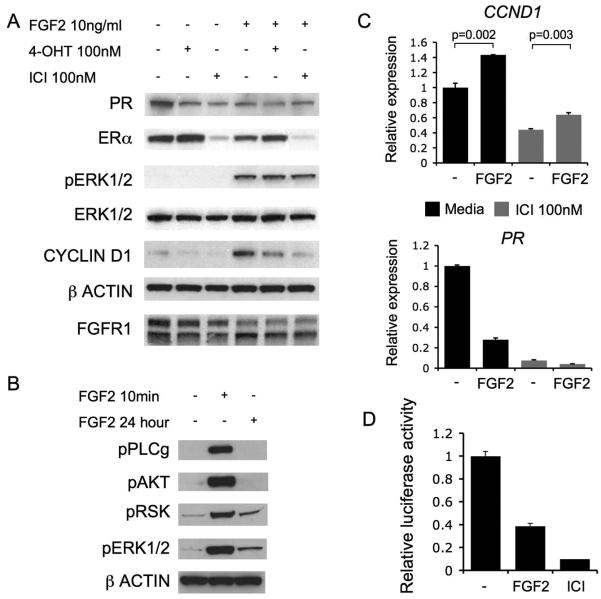
Figure 5. Signalling in SUM44 cells is response to endocrine therapies.
A. Western blots of PR, ER, phosphorylated-ERK1/2, ERK1/2, CCND1, β-ACTIN, and FGFR1. SUM44 cell lysates treated for 24 hours prior to lysis with 100nM 4-OHT, 100nM ICI-182780, or no treatment (−), with or without 10ng/ml FGF2. Phosphorylated-AKT1 was not detected.
B. Western blots of phosphorylated-PLCγ1 (Tyr783), phosphorylated-AKT, phosphorylated-p90RSK (Thr359/Ser363), phosphorylated-ERK1/2, and β-ACTIN on SUM44 cell lysates treated for either 10 minutes or 24 hours with 10ng/ml FGF2 prior to lysis.
C. Quantitative RT-PCR analysis of cyclin D1 (CCND1 - Top) and (PR - Bottom) expression in SUM44 cells treated with or without 10ng/ml FGF2 for 24 hours prior to RNA isolation, without (Black bars) or in the presence of 100nM ICI-182780 (Grey bars).
D. SUM44 cells were co-transfected with EREIItkLuc (ERE-luciferase reporter construct) and pCH110 (β-galactosidase reporter construct) and treated for 48 hours with 10ng/ml FGF2, or no treatment, with 100nM ICI-182780 as positive control. Luciferase activity was expressed relative to β-galactosidase activity. Error bars SEM of 3 repeats, p values Student's T-test.

